
Prerequisites for High-Resolution Molecular Mapping
Executing a high-resolution proteomics workflow requires a departure from the traditional, static view of disease progression. Practitioners must abandon the notion that rare diseases move through discrete, linear stages. Instead, the objective is to reconstruct a molecular continuum. For instance, the progression of Metabolic dysfunction-associated steatotic liver disease (MASLD) is no longer viewed as a simple jump from steatosis to cirrhosis, but as a continuous trajectory of regulatory programs and cellular remodeling. To map this, the analyst requires high-fidelity access to both tissue-specific transcriptomic profiles and paired plasma proteomics to identify markers that are actually accessible in a clinical setting.
- Unbiased mass spectrometry-based proteomics for cerebrospinal fluid (CSF) or plasma analysis.
- Single-cell RNA sequencing (scRNA-seq) to identify cell-type-specific signatures across diverse populations.
- Integrated transcriptomic-proteomic datasets to validate plasma-accessible biomarker panels.
- High-resolution spatio-temporal activity mapping tools for neurological targets, such as those used in dorsal raphe nucleus (DRN) studies.
Executing the Proteomic Workflow
- Sample Acquisition: Secure paired samples (e.g., liver tissue and plasma or CSF and blood) to ensure the molecular signals found in the organ are mirrored in accessible biofluids.
- Unbiased Protein Identification: Employ mass spectrometry to detect coordinated increases in specific protein families, such as the HDL-like lipid transport proteins APOA1, APOA2, and APOL1 observed in neurodegenerative contexts.
- Signature Filtering: Isolate cell-type-specific clusters—such as GPNMB+ and CD74+ microglial subgroups—that remain consistent across multiple ethnic population groups.
- Trajectory Reconstruction: Use data-driven frameworks to position patients along a continuous molecular trajectory rather than assigning them to a rigid clinical stage.
- Validation of Circuitry: Confirm that identified proteins interact with known transcriptional drivers, such as the Rbm5-Myc interaction in leukemia stem cells, to ensure the biomarker is functional and not just correlative.
The precision of this workflow is best illustrated by the Phase 2 FLICKER study. By utilizing unbiased, mass spectrometry-based proteomics on cerebrospinal fluid, investigators identified that sensory stimulation via the Spectris platform directly increased levels of HDL-like lipid transport proteins. This wasn't a random fluctuation; it was a coordinated modulation of myelination pathways. When you target the CSF, you are looking at the immediate biochemical environment of the brain, allowing for the detection of APOA1 and APOA2 increases that would be diluted or invisible in a standard systemic blood test.

Integrating this with a data-driven framework allows for the creation of highly predictive biomarker panels. In the case of MASLD, the integration of liver-plasma proteomics led to the identification of a 57-gene plasma-accessible biomarker panel. This panel does more than predict advanced fibrosis; it continuously positions the patient along the disease trajectory. Why does this matter? Because conventional non-invasive clinical scores often fail to capture the nuance of early-stage cellular remodeling, whereas a 57-gene proteomic signature captures the actual molecular state of the liver.
| Metric | Conventional Stage-Based Model | High-Resolution Molecular Continuum |
|---|---|---|
| Patient Stratification | Discrete Stages (e.g., Fibrosis Stage 1-4) | Continuous Molecular Trajectory |
| Biomarker Precision | General clinical scores | 57-gene plasma-accessible panel |
| Analysis Depth | Symptomatic observation | Cell-type specific signatures (e.g., SST+ GABAergic) |
| Population Scope | Often single-cohort | Cross-population shared signatures |
The complexity of rare diseases often stems from their heterogeneity across different populations. High-resolution workflows must account for this by identifying signatures that are shared across ethnic groups. In Alzheimer's disease research, this involves isolating specific cell-type signatures—such as SERPINH1+, CD44+, and WIF1+ astrocytic subgroups—that persist regardless of the patient's background. By focusing on these shared molecular subtypes, researchers can develop therapies that are universally applicable rather than cohort-specific.
"The transition from stage-based classification to a continuous molecular trajectory allows us to resolve the ordered activation of regulatory programs that were previously invisible."— Nature Research, MASLD Framework Analysis
Beyond systemic diseases, mapping rare disorders requires understanding the specific transcriptional circuitry that sustains pathology. Take Acute Myeloid Leukemia (AML) as an example. The RNA-binding protein Rbm5 has been identified as a critical sustainer of leukemia stem cells (LSCs). Through single-cell RNA sequencing, it became clear that Rbm5 loss specifically decreases the proportion of LSCs by downregulating the stemness gene Myc. The proteomics workflow here isn't just about counting proteins; it is about mapping the physical interaction between RBM5 and MYC to uncover the autoregulatory mechanism driving the malignancy.
Targeted Protein Restoration
For ultra-rare neurological disorders like Dravet syndrome, the goal is the restoration of specific protein production. ION337, an RNA-targeted medicine, focuses on increasing the production of the NaV1.1 protein, which is deficient due to SCN1A gene variants. The technical objective here is high potency with infrequent intrathecal dosing—potentially every six months.
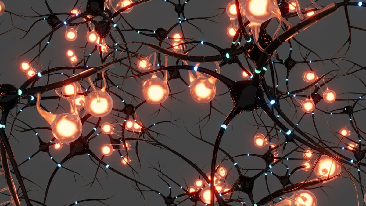
Finally, the frontier of mapping involves the spatiotemporal organization of activity. In zebrafish models of the dorsal raphe nucleus (DRN), researchers found that neurons are topographically organized to target specific forebrain regions. This means that sensory-motor responses are not global but are mapped to specific coordinates. When DRN neurons are ablated, the synchrony of forebrain neurons collapses, enhancing defensive behaviors. This level of resolution—mapping the protein-driven activity of specific axons—is the gold standard for understanding how rare neurological deficits manifest as behavioral changes.
Common Pitfalls in Proteomic Mapping
Most failures in rare disease mapping occur when researchers rely on outdated clinical scoring systems that assume a linear progression. These systems ignore the molecular continuum and fail to account for the fact that two patients in the same clinical stage may have entirely different molecular trajectories. Furthermore, ignoring the cell-type specificity of a protein signature can lead to false positives; a protein increase in a microglial subgroup may have a completely different pathological meaning than the same increase in a neuronal subgroup.
- Over-reliance on aggregate tissue proteomics instead of single-cell resolution.
- Failure to validate plasma-accessible markers against tissue-specific transcriptomes.
- Ignoring the role of RNA-binding proteins (like Rbm5) in maintaining stem cell self-renewal.
- Neglecting the impact of population-specific genetic variance on shared cell-type signatures.
To avoid these traps, the practitioner must ensure that every identified biomarker is cross-referenced with a functional circuit. If a protein like APOA1 is increased, the analyst must ask if it correlates with a specific pathway—such as lipid transport or myelination—and if that pathway is consistently modulated across the study cohort. Only by linking the proteomic signal to a biological mechanism can we move from simple observation to actionable therapeutic design.