
Prerequisites for Clinical Readiness
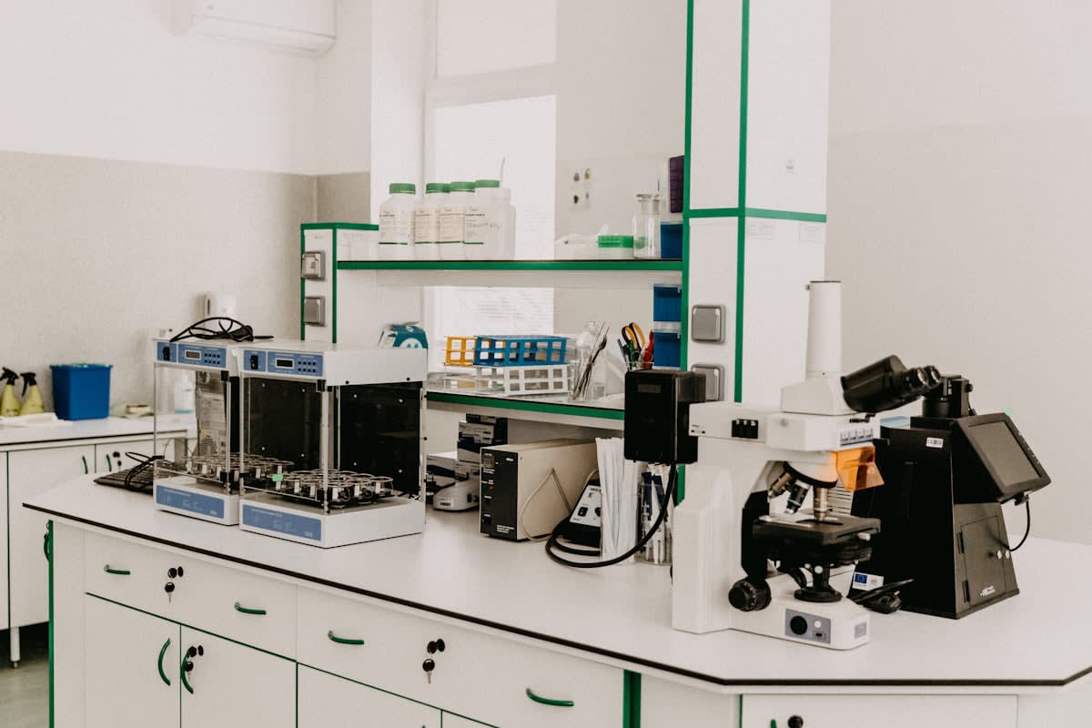
Implementing an autologous stem cell program requires more than just a skilled clinician; it demands a rigorous architectural shift in the clinic's operational environment. The primary objective is the mitigation of the processing gap—the critical window between cellular harvest and delivery where viability often plummets. To achieve clinical consistency, a facility must establish a controlled environment that adheres to ISO 7 or ISO 5 cleanroom standards for any open-system processing. This ensures that the cellular concentrate remains free from exogenous contaminants that could trigger an inflammatory response or jeopardize patient safety. Without this foundational infrastructure, the variance in patient outcomes becomes an unpredictable variable rather than a manageable clinical metric.
- Closed-system centrifugation units with programmable G-force and temperature control.
- Sterile, single-use aspiration and processing kits to eliminate cross-contamination.
- High-resolution imaging (Ultrasound or Fluoroscopy) for precise delivery.
- Hemocytometers or automated cell counters for quantitative viability assessment.
- Regulatory certifications compliant with FDA (US), EMA (EU), or PMDA (Japan) guidelines.
Staffing requirements must move beyond general nursing to include technicians trained in aseptic processing and cellular handling. The precision of the centrifuge cycle, for instance, can be the difference between a high-yield mesenchymal stem cell (MSC) concentrate and a useless slurry of red blood cells. Practitioners must implement a double-verification system for all cellular products, ensuring that the identity of the donor and the volume of the concentrate are logged before the first injection is administered. This level of rigor transforms a boutique offering into a scalable, medical-grade protocol.
The Implementation Protocol
The transition from patient intake to cellular delivery must follow a linear, non-deviant path. Many clinics fail because they treat the procedure as a single event rather than a multi-stage biological process. By decoupling the harvest from the delivery, clinicians can perform quality control checks that ensure the patient is receiving a therapeutic dose of cells, not just a volume of fluid. This systematic approach is what separates high-performance regenerative centers from those relying on anecdotal evidence.
- Patient Biomarker Screening and Selection
- Targeted Cellular Harvesting (BMAC or SVF)
- Controlled Centrifugation and Processing
- Quantitative Viability and Concentration Analysis
- Precision-Guided Delivery
- Post-Procedural Biological Monitoring
Patient selection begins with a biomarker profile to determine the likely yield of the harvest. Not all patients are equal candidates; age, systemic inflammation, and comorbidities like Type 2 diabetes can significantly reduce the concentration of MSCs in the bone marrow or adipose tissue. For example, clinicians in Southeast Asian hubs have noted that metabolic health correlates strongly with the proliferative capacity of the harvested cells. A pre-procedure assessment of C-reactive protein (CRP) levels can help manage patient expectations and adjust the harvest volume to ensure a therapeutic threshold is met.
Harvesting techniques vary based on the target pathology. Bone Marrow Aspirate Concentrate (BMAC) is typically preferred for orthopedic applications due to its rich concentration of hematopoietic and mesenchymal cells. Conversely, Stromal Vascular Fraction (SVF) derived from adipose tissue offers a higher absolute number of MSCs but requires a more complex enzymatic digestion process. The choice between these two is not merely a matter of preference but of biological requirement; the different secretomes of BMAC and SVF trigger distinct healing cascades in the recipient tissue.
| Feature | BMAC (Bone Marrow) | SVF (Adipose) |
|---|---|---|
| Cell Yield | Low to Moderate | High |
| Processing Complexity | Low (Centrifugation) | High (Digestion/Centrifugation) |
| Primary Indication | Joint/Bone Repair | Soft Tissue/Aesthetics |
| Typical MSC Concentration | 0.001% - 0.01% | 0.1% - 1% |
Processing is where most clinical errors occur. The use of excessive G-force during centrifugation can lead to cell lysis or mechanical stress that triggers premature senescence. A standardized protocol should utilize a two-stage spin: a low-speed spin to separate the buffy coat from the plasma and red cells, followed by a precise concentration step. Maintaining the temperature at 4 degrees Celsius during this process is non-negotiable to prevent metabolic exhaustion of the cells before they reach the target site.
Once processed, the concentrate must be quantified. Relying on the visual appearance of the buffy coat is an amateur mistake. Using a hemocytometer to determine the total nucleated cell (TNC) count allows the practitioner to calculate the exact dosage. If the TNC falls below a predetermined threshold—typically 10-20 million cells per mL for orthopedic applications—the clinician must decide whether to proceed or repeat the harvest. This quantitative data provides the only real basis for comparing outcomes across a patient cohort.
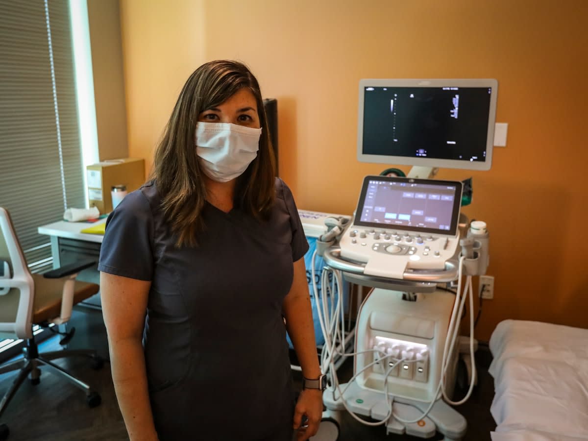
Delivery is the final critical link. Injecting stem cells blindly into a joint or tissue is an inefficient use of biological material. The use of fluoroscopy or high-resolution ultrasound allows the clinician to place the concentrate precisely within the target lesion. This localized delivery maximizes the interaction between the MSCs and the damaged tissue's signaling molecules. Furthermore, the injection speed must be controlled; rapid bolus injections can create hydrostatic pressure that may damage the fragile cell membranes of the concentrate.
Optimizing Cellular Viability
Viability is the only metric that truly matters in regenerative medicine. A high cell count is meaningless if the cells are non-viable or functionally dormant. To optimize this, clinics must scrutinize their reagents. The transition to animal-free, xeno-free processing media reduces the risk of immunogenic reactions and improves the stability of the cells. Moreover, the time from harvest to injection should be kept under 60 minutes. Every ten minutes of delay at room temperature can result in a measurable drop in the metabolic activity of the MSCs.
"The difference between a failed regenerative procedure and a successful one is rarely the cells themselves, but the precision of the processing pipeline that delivers them."— Dr. Aris Thorne, Regenerative Systems Analyst
Regulatory Safeguard
Ensure that all processing steps are documented in a cellular chain-of-custody log. In the event of an adverse reaction or a regulatory audit, the ability to trace the exact G-force, temperature, and TNC count is the only legal and clinical defense.
Critical Failures and Common Pitfalls
Contamination remains the most catastrophic failure mode. This often occurs not during the harvest, but during the transfer of the concentrate from the centrifuge tube to the injection syringe. Using open-system transfers in a non-sterile environment introduces opportunistic pathogens that can lead to septic arthritis or systemic infection. The implementation of a fully closed-loop system, where the cells never encounter ambient air, is the only way to virtually eliminate this risk.
Another frequent error is the 'volume fallacy'—the assumption that more volume equals more healing. In reality, over-diluting the concentrate to fill a larger joint space can lower the local concentration of signaling molecules below the therapeutic threshold. Practitioners must focus on the cell density (cells per microliter) rather than the total volume of the injection. A concentrated 2mL dose is vastly superior to a diluted 10mL dose that fails to trigger the necessary regenerative cascade.
Finally, the lack of standardized post-procedural protocols often masks the true efficacy of the treatment. Many clinics fail to prescribe a specific loading or rehabilitation phase that encourages the newly injected cells to differentiate and integrate into the tissue. Without a controlled mechanical stimulus, MSCs may remain quiescent or be resorbed by the body without providing a structural benefit. A blueprint for implementation is incomplete without a corresponding blueprint for post-injection biological activation.