Indian scientists produce most detailed 3D atlas of the human brainstem
Source Entity
Hacker News
<a href="https://news.ycombinator.com/item?id=48903082">Comments</a>
A Landmark Achievement in Neuroanatomy
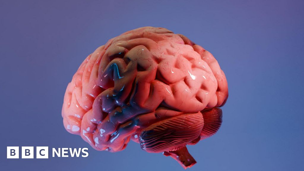
Indian scientists have successfully developed the most detailed 3D atlas of the human brainstem to date. This achievement represents a significant leap forward in our understanding of one of the most complex and least accessible regions of the human central nervous system. The brainstem, which serves as the critical conduit between the cerebrum, cerebellum, and the spinal cord, is responsible for regulating fundamental autonomic functions such as breathing, heart rate, and blood pressure. By producing a high-resolution three-dimensional map, these researchers have provided the global scientific community with an unprecedented tool for studying the structural organization of the human brain.
The Technical Significance of a 3D Atlas
Creating a 3D atlas of the brainstem is an immense technical challenge due to the region's dense packing of nuclei and intersecting nerve tracts. Unlike the cerebral cortex, where structures are more spread out, the brainstem is a compact hub of vital circuitry. The precision of this new atlas allows for the exact localization of specific nuclei and pathways that were previously blurred or poorly defined in lower-resolution models. This level of detail is crucial for neurosurgeons and neurologists who must navigate this high-stakes area where even a millimeter of deviation during a procedure can lead to catastrophic loss of motor function or respiratory failure.
Broader Implications for Neurological Medicine
The implications of this mapping effort extend far beyond basic anatomy. Many devastating neurological conditions, including brainstem strokes, gliomas, and degenerative diseases like Amyotrophic Lateral Sclerosis (ALS), originate or manifest within this region. With a more accurate 3D map, clinicians can better predict how a lesion or tumor will affect specific bodily functions based on its precise spatial coordinates. Furthermore, this atlas provides a baseline for comparing healthy brainstem architecture against pathological states, potentially leading to earlier diagnosis and more targeted therapeutic interventions for patients suffering from brainstem-related disorders.
Historical Context and the Evolution of Brain Mapping
Historically, brain mapping relied on two-dimensional histological slices, which required researchers to mentally reconstruct 3D structures—a process prone to error and inconsistency. The transition to 3D imaging, powered by advancements in Magnetic Resonance Imaging (MRI) and computational reconstruction, has revolutionized the field. This Indian-led initiative builds upon decades of global efforts to map the human connectome, but it distinguishes itself by focusing specifically on the brainstem's intricate architecture. It signals a shift toward high-precision anatomical research in India, leveraging cutting-edge imaging technology to contribute to foundational global science.
Future Trends: AI and Personalized Neuro-Mapping
Looking ahead, this 3D atlas is likely to serve as a training dataset for artificial intelligence and machine learning algorithms. AI can be trained on these detailed maps to automatically identify anomalies in patient scans, drastically reducing the time required for radiological analysis. We are moving toward an era of 'personalized neuro-mapping,' where a patient's individual brainstem structure can be overlaid onto this gold-standard atlas to create a customized surgical plan. This integration of big data and anatomy will likely lead to a new generation of minimally invasive neurological surgeries with significantly higher success rates.
Conclusion
The production of the most detailed 3D atlas of the human brainstem by Indian scientists is more than just a mapping exercise; it is a foundational contribution to medical science. By illuminating the hidden corridors of the brainstem, this work paves the way for safer surgeries, deeper insights into autonomic failures, and a more sophisticated understanding of human consciousness and survival. As this data is integrated into clinical practice and AI-driven diagnostics, the impact will be felt in hospitals and research labs worldwide.